

Takeaway
A family history is not a diagnosis. It is evidence that something specific runs through your family, and specific things can be found if you look with the right instrument.
For most people in your position the finding falls into one of three categories. An inherited particle that no routine panel screens for. A particle count that a normal LDL concealed. Or a metabolic problem that appeared in insulin years before it would have appeared in glucose. Each calls for a different response, and none of them show up on the test you have been getting every year.
Your father had a stent at 52. Your grandfather never made it past 60. And every year at your physical, the doctor looks at your cholesterol panel, says it looks fine, and moves on to the next thing.
A standard lipid panel measures four values total cholesterol, LDL, HDL, and triglycerides. It was built as a population screening tool and it does that job reasonably well. What it was never built to do is explain why cardiovascular disease keeps appearing in one particular family. Having a first-degree relative with early heart disease raises your risk independently of your cholesterol numbers, which is another way of saying the standard panel measures something other than what is going wrong in your bloodline.
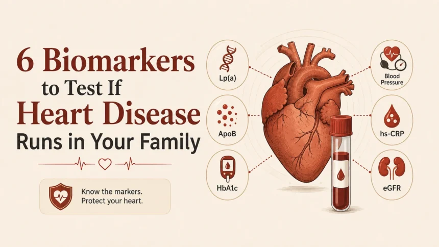
Six blood markers cover most of what it misses. They are not equally important. One of them matters more than the other five put together, so treat this as a priority list rather than a checklist.
Lipoprotein(A): The Test Almost Nobody Has Had
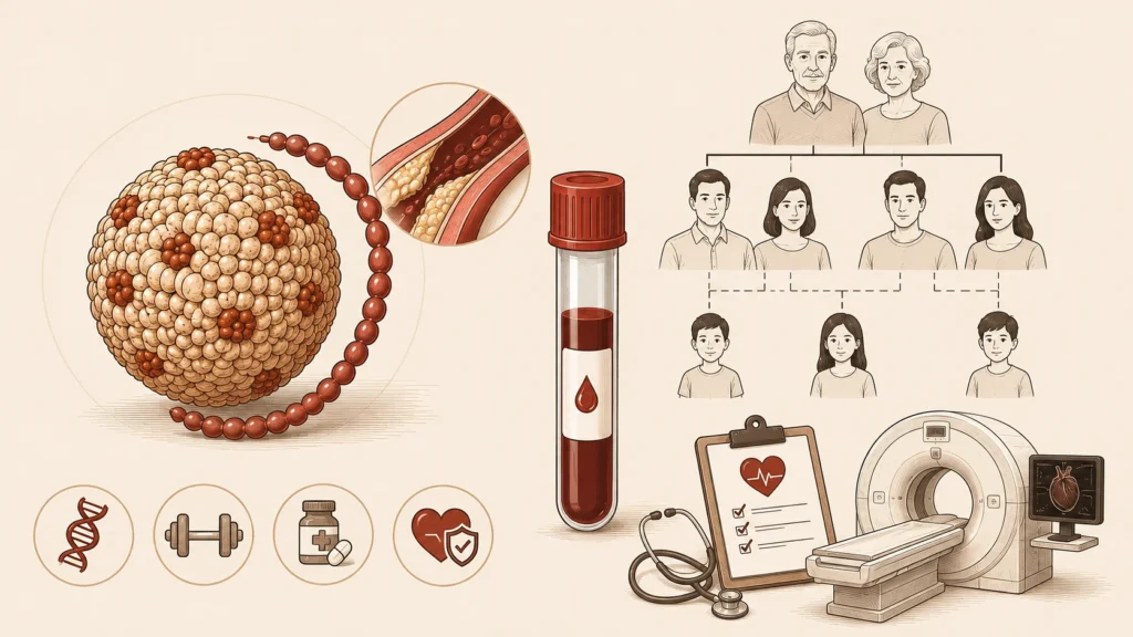
Lp(a) is an LDL particle with an extra protein attached to it. Your level is fixed at conception. Diet will not change it. Exercise will not change it. Statins do not lower it and in some people raise it slightly. Whatever number you were born with is the number you will still have at 70.
Roughly one person in five carries an elevated level. Most of them never learn this, because Lp(a) appears on no routine panel and factors into no standard risk calculator. If heart disease runs through your family and nobody’s cholesterol ever looked bad enough to explain it, start here. This is the most common inherited driver of early cardiovascular disease and also the most frequently missed one.
Why It Does More Damage Than Its Numbers Suggest
The mechanism works on two fronts at once. The LDL-like core deposits cholesterol into artery walls the way any LDL particle does. Meanwhile the attached apo(a) protein closely resembles plasminogen, which is the enzyme your body uses to dissolve clots. It resembles it closely enough to interfere with it.
So elevated Lp(a) speeds up plaque formation while simultaneously weakening your ability to break down a clot once one forms. That pairing explains why it produces events at younger ages than lipid burden alone would predict.
What A High Result Actually Changes
Your treatment targets tighten. No approved therapy lowers Lp(a) directly. Several are in late-stage trials, none have reached the market. The clinical response is therefore to push harder on everything that is modifiable. LDL targets drop. Blood pressure control gets stricter. Statin therapy may begin ten years earlier than your other numbers alone would warrant.
Your family needs testing. Lp(a) is inherited codominantly. Your siblings have roughly a 50% chance of carrying the same elevation, and your children a similar chance. One result on you produces actionable information for four or five other people. Very few blood tests do that.
Imaging becomes reasonable. High Lp(a) combined with family history makes a strong case for a coronary artery calcium scan. The question stops being what your statistical risk is and becomes whether there is plaque in your arteries right now. A CAC scan answers that.
Apolipoprotein B: Counting Particles Instead Of Contents
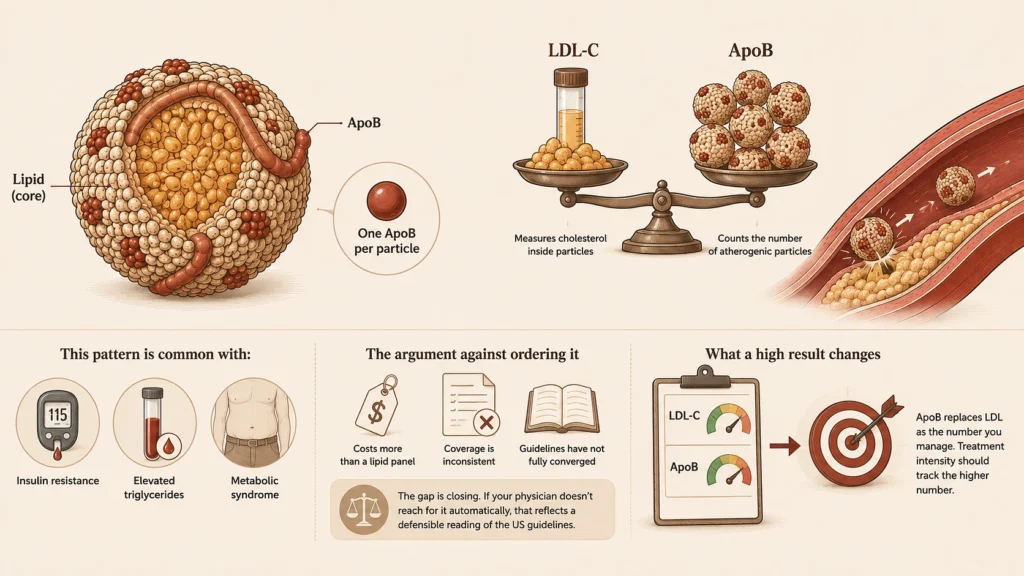
LDL cholesterol tells you how much cholesterol is being carried inside lipoprotein particles. ApoB tells you how many particles there are. Every atherogenic particle carries exactly one ApoB molecule, so the count comes out direct rather than estimated.
Most of the time the distinction stays invisible, because in most people the two numbers agree. In a sizable minority they do not, and when they disagree, ApoB predicts events better. Arterial damage depends on how many particles collide with the artery wall and lodge there, not on the total cholesterol those particles happen to be carrying. Someone whose particles are small and cholesterol-poor can post a comfortable LDL while carrying a particle count in a high percentile.
That pattern is not unusual. It shows up with insulin resistance, elevated triglycerides, and metabolic syndrome, meaning it is most common in precisely the people whose physical ends with the phrase "your cholesterol is fine."The Argument Against Ordering It
ApoB costs more than a lipid panel, coverage is inconsistent, and the major guidelines have not fully converged. European guidance treats it as the preferred measure in patients with high triglycerides, diabetes, obesity, or very low LDL. American guidance has stayed more conservative, positioning it as a risk-refining measurement rather than a primary treatment target.
The gap is closing. But if your physician does not reach for it automatically, that reflects a defensible reading of the US guidelines rather than negligence.
What a high result changes: ApoB replaces LDL as the number you manage. If your LDL looks acceptable and your ApoB does not, you have located the thing the standard panel was hiding, and treatment intensity should track the higher number rather than the more comfortable one.
Fasting Insulin: The Years Before Glucose Moves
Glucose is a lagging indicator. By the time fasting glucose or HbA1c drifts out of range, the pancreas has usually been compensating for years, secreting steadily more insulin to hold blood sugar in place. Those years are exactly when intervention works best, and standard testing passes straight through them without registering anything.
Fasting insulin measures the compensation itself. It is the earliest routinely available signal that metabolic machinery is under strain.
This ties directly back to ApoB. Insulin resistance produces a consistent lipid signature: triglycerides rise, HDL falls, and LDL particles shift toward smaller and denser. That combination generates a high particle count sitting behind a normal-looking LDL. Elevated fasting insulin is frequently the upstream explanation for an ApoB result that does not match the lipid panel.
A Warning About Reference Ranges
Lab reference ranges for insulin are considerably wider than the physiology justifies. Many labs flag a result only above 25 µIU/mL, a threshold drawn from population distributions in a population where insulin resistance is widespread. Clinicians who use this marker regularly tend to treat single digits as normal and anything in the teens as worth addressing.
Assay standardization across labs is also imperfect. Track your trend within one lab rather than comparing results between two.
What a high result changes: the intervention moves upstream. Rather than managing lipids with medication, you address carbohydrate load, resistance training, sleep, and body composition. Lipid abnormalities frequently improve as a downstream consequence. Of everything on this list, this is the marker where the primary response is behavioral rather than pharmaceutical.
High-Sensitivity CRP: Measuring Plaque Stability
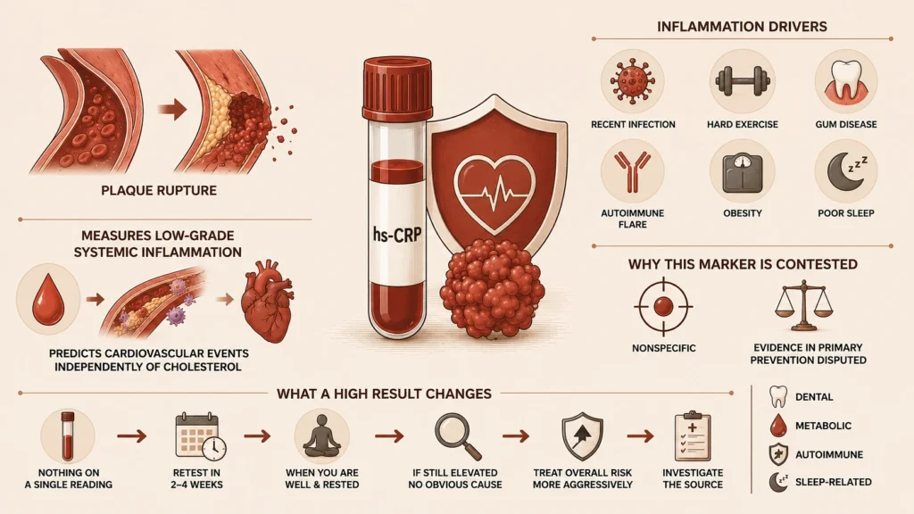
Most heart attacks do not come from an artery narrowing gradually until it closes. They come from an unstable plaque rupturing, with a clot forming across the tear within seconds. Inflammation is what destabilizes plaque, degrading the fibrous cap that holds its contents in place.
hs-CRP measures low-grade systemic inflammation and predicts events independently of cholesterol. The high-sensitivity assay matters here. Standard CRP is calibrated to detect the large spikes of active infection and cannot resolve the subtle range that carries cardiovascular meaning.
Why This Marker Is Genuinely Contested
hs-CRP is nonspecific, and that is a real limitation rather than a footnote. A recent cold raises it. So does a hard training session, gum disease, an autoimmune flare, obesity, or a stretch of poor sleep.
Its evidentiary weight in primary prevention is also disputed. The US Preventive Services Task Force has repeatedly declined to endorse routine hs-CRP screening, arguing it has not been shown to reclassify enough people into different treatment categories to justify testing everyone. That position is reasonable. The case for testing it is stronger in your situation than in the general population, because family history already places you in the range where reclassification carries the most consequence.
What a high result changes: nothing, on a single reading. Retest in two to four weeks when you are well and rested. If it stays elevated with no obvious cause, it argues for treating your overall risk more aggressively and for investigating the source. Dental, metabolic, autoimmune, and sleep-related causes are the usual suspects.
Cystatin C: Kidney Function Without The Muscle Problem
Kidney function ranks among the strongest predictors of cardiovascular mortality, and the standard way of measuring it carries a structural flaw.
Creatinine is a byproduct of muscle breakdown. Estimated GFR calculated from it therefore depends on how much muscle you carry. A muscular person can appear to have reduced kidney function they do not have. An older or sarcopenic person can appear normal while their function is meaningfully impaired. The second error is the dangerous one, because nothing about it is visible.
Cystatin C is produced by essentially all nucleated cells at a constant rate and does not track muscle mass. Estimates combining cystatin C with creatinine outperform either alone, and current kidney guidelines recommend cystatin C for confirming a borderline creatinine result.
What a high result changes: reduced kidney function raises cardiovascular risk on its own and alters prescribing, since several relevant medications require dose adjustment or avoidance below certain thresholds.
Lp-PLA2: Included For Completeness, Not Recommended
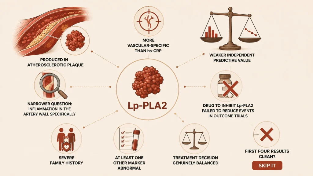
Lipoprotein-associated phospholipase A2 is produced inside atherosclerotic plaque, which makes it more vascular-specific than hs-CRP. In principle it answers a narrower question: whether inflammatory activity is happening in the artery wall specifically, rather than somewhere in the body generally.
In practice, its independent predictive value has looked weaker in large pooled analyses than early studies suggested. The drug developed to inhibit the enzyme failed to reduce events in outcome trials, which undercut the case that it drives disease rather than simply accompanying it.
It earns a place only when family history is severe, at least one other marker is abnormal, and a treatment decision sits genuinely balanced. If your first four results come back clean, skip it.How To Order The Panel
Start with Lp(a), ApoB, hs-CRP, and fasting insulin. Those four cover distinct pathways: inherited particle burden, total particle count, vascular inflammation, and metabolic dysfunction. Any one of them can be the answer on its own.
Add cystatin C if you are over 50 or have any prior kidney or blood pressure concern. Add Lp-PLA2 only if the first round comes back abnormal.
Practical notes:
- Fast eight to twelve hours. Fasting insulin and triglycerides are meaningless otherwise.
- Do not test within two weeks of an illness or a heavy training block.
- Repeat any elevated inflammatory marker before drawing conclusions from it.
- Take the results to a physician who will act on them. Ordering markers your doctor will not interpret produces anxiety rather than care.